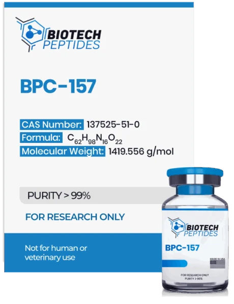
Buy BPC-157 (5mg & 10mg) Online
Buy BPC-157 (5mg & 10mg) Online. BPC-157, Body Protection Compound-157, is obtained from the parent protein Body Protection Compound (BPC). BPC is a naturally occurring protein in the digestive tract.[1] BPC -57 is a penta-decapeptide made up of 15 amino acids, and is derived from a stretch of endogenous BPC identified and isolated from gastric juice. Animal studies have suggested its potential in supporting tissue repair processes in muscle, tendon, and torn ligaments. It may further protect organs and potentially prevent gastric ulcer development.[2] Sikiric et al. noted that there was “a strong protection, noted following [exposure to] BPC 157.” BPC-157 also has the potential to enhance the function of the digestive tract and prevent against irritable bowel syndrome (IBS), gastrointestinal cramps, and Crohn’s disease. The peptide also has possible analgesic characteristics.
Specifications
Molecular Formula: C62H98N16O22
Molecular Weight: 1419.556 g/mol
Sequence: L-Valine,glycyl-L-alpha-glutamyl-L-prolyl-L-prolyl-Lprolylglycyl-L-lysyl-L-prolyl-L-alanyl-L-alpha-aspartyl-L-alpha-aspartyl-L-alanylglycyl-L-leucyl-;glycyl-L-alpha-glutamyl-L-prolyl-L-prolyl-L-prolylglycyllysyl-L-prolyl-L-alanyl-L- alpha-aspartylL-alpha-aspartyl-L-alanylglycyl-L-leucyl-L-valine
BPC-157 Peptide Research
BPC-157 and Wound Healing
The GI tract’s mucosal barrier is considered to protect the underlying tissues from the harmful actions of bile, gastric acid, and other compounds necessary for the digestion and absorption of nutrients from food. BPC-157 is believed to help preserve the structural integrity of the mucosal layer. The role appears to be partially mediated through the recruitment of fibroblasts. Fibroblasts are considered to produce extracellular matrix proteins such as fibrin, collagen, elastin, and others. BPC-157 has been suggested to promote the proliferation and faster migration of fibroblasts in a concentration-dependent manner.[3] In another research study it was hypothesized that BPC-157 may have led to an acceleration in wound closure compared to the control group via an improvement in the formation of granulation tissue, reepithelialization, dermal remodeling, and collagen deposition. There is a possibility that BPC-157 may have promoted the expression of vascular endothelial growth factor (VEGF) in the injured skin tissues.[4] Moreover, the researchers commented that BPC-157 may have shown a potential to enhance umbilical vein endothelial cell proliferation (HUVECs). Furthermore, there may have been a significant increase in the migration of HUVECs, as indicated by the comments from wound healing assays. BPC-157 possibly resulted in an upregulation of VEGF-a expression and acceleration of vascular tube formation. It also appeared that BPC-157 may have regulated the phosphorylation level of extracellular signal-regulated kinases 1 and 2 (ERK1/2) and their downstream targets, including c-Fos, c-Jun, and Egr-1. These molecules are hypothesized to potentially play significant roles in cell growth, migration, and angiogenesis.[4]
BPC-157 and Vascular Growth and Collateralization
The peptide has the potential as an angiogenic, and studies suggest it may enhance endothelial cells’ growth and proliferation, which line the walls of blood vessels. Research in rats has observed that the peptide may substantially increase the collateral blood vessel growth rate in the setting of ischemia.[5] That action has been primarily observed in the GI tract, but research has noted similar observations in muscle, neurological, and cardiovascular tissues. Research using chicken embryos has posited that BPC-157 may also have the potential to promote vascular growth through activation of VEGFR2 pathway. VEGFR2 is a cell surface receptor active in nitric oxide signaling and is considered to support cell activity and proliferation. BPC-157 may promote vascular “running” in cultured cells. This is the growth and development of new blood vessels towards a site of injury or around the area of blood clot to reach out to distal tissues and thus protect cellular function.
BPC-157 and Tendon Healing
BPC-157 studies have observed potential in connective tissue healing such as ligament, bone, and tendon. Ligament and tendon injuries take a long time to heal due to the poor blood supply to these tissues. There is slower migration of fibroblasts and wound-healing cells to these sites of injury owing to the poor blood supply, and therefore the repair process is obstructed. The peptide has the potential to improve the collateralization and density of fibroblasts in the sites of injury in research involving rat tendons.[6] More specifically, the study also hypothesized that BPC-157 might accelerate the outgrowth of tendon fibroblasts from tendon explants. This suggests that BPC-157 may potentially promote the growth of new cells in the injured tendon. Further, the survival of the cells to which BPC-157 was applied may be significantly increased when exposed to H(2)O(2) stress, indicating a potential protective action against oxidative stress. BPC-157 may also enhance the migration of tendon fibroblasts, as indicated by the transwell filter migration assay used in the study. This may imply that BPC-157 potentially promotes the movement of tendon fibroblasts. Moreover, BPC-157 may accelerate the spreading of tendon fibroblasts on culture dishes, suggesting potentially increased cell adhesion and attachment.[6] This experimental research has suggested BPC-157 to be a positive impactor in comparison to EFG, bFGF, and VGF hormones. Immunostaining assays involving FITC conjugated phalloidin have suggested BPC-157 to enhance F-actin formation in fibroblasts. F-actin is considered to be crucial for cell structure and function and promotes cell migration. Immunoblotting experiments have noted that BPC-157 appears to increase the phosphorylation of paxillin and FAK proteins, which are considered crucial for cellular migration. More specifically, BPC-157 may induce F-actin formation in tendon fibroblasts, potentially indicating enhanced cytoskeletal organization and cell motility. Further analysis using Western blotting indicates that BPC-157 may activate the FAK (focal adhesion kinase) and paxillin proteins. The phosphorylation levels of FAK and paxillin may increase with BPC-157, while the total amounts of these proteins may remain unchanged. This potentially suggests that BPC-157 may activate the FAK-paxillin pathway, which may be involved in the promotion of tendon fibroblast migration and cell adhesion
Be the first to review “BPC-157 (5mg & 10mg)”
Related products
Peptides
Peptides
Peptides
Peptides
Peptides
Peptides
Peptides
Peptides
Reviews
There are no reviews yet.